In 1895, Wilhelm Röntgen captured an extraordinary image: an X-ray of his wife’s hand. Her bones and wedding ring were visible, offering a striking demonstration of a new way to look inside the living body.
That early image was not a cancer scan. Yet it helped open a path towards modern cancer imaging: the techniques clinicians use to investigate suspicious findings, assess the extent of disease and follow changes during treatment. [1]
More than a century later, that view inside the body has become much richer. Each technology contributes something different, and each still depends on careful interpretation.
X-rays and the First Window Inside
X-rays quickly became useful in medicine because they could show structures that had previously been hidden. Bones appear clearly, but many soft tissues are harder to distinguish on a conventional X-ray.
Specialised techniques developed to answer more specific questions. Mammography, for example, uses X-rays to examine breast tissue and remains an important screening tool. The development of newer scanners has not made every earlier technique obsolete; the clinical question determines which test is useful.
Ultrasound and Imaging with Sound
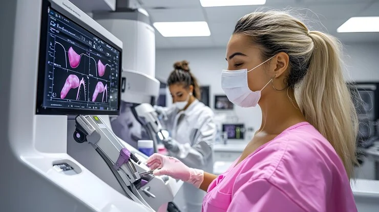
Ultrasound uses high-frequency sound waves to form images in real time, without ionising radiation. It can help clinicians examine organs, assess certain lumps and lymph nodes, and guide needles during biopsies or other procedures.
It offers a different perspective from X-rays and is particularly useful when an examination needs to follow movement or guide a procedure as it happens. It is one part of the imaging toolkit, rather than a replacement for every other scan. [2]
CT and the Body in Cross Section
The arrival of clinical CT in the 1970s changed what doctors could see. By combining X-ray measurements taken from different angles, a computer could reconstruct cross-sectional views instead of a single overlapping projection. [1]
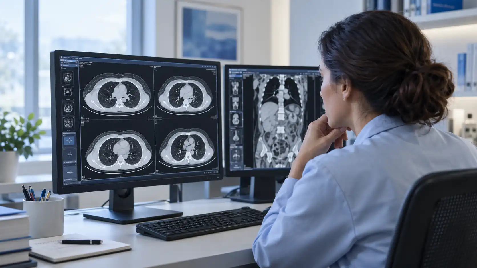
In cancer care, CT can help locate a tumour, assess its size and look for possible spread. Comparing scans over time can help the treating team evaluate changes during treatment. CT is also used in planning or guiding some procedures. [3]
CT uses ionising radiation. The decision to scan should weigh the expected clinical benefit against its risks, rather than assume that more imaging is always better. [3]
MRI and Soft Tissue Detail
MRI uses magnetic fields and radio waves rather than ionising radiation. Its ability to distinguish soft tissues can help clinicians examine a tumour and its relationship to nearby structures. [2]
Brain, breast, liver, prostate, rectal and musculoskeletal cancers are among the areas where MRI can contribute valuable information. The appropriate scan and protocol depend on the organ and the question being asked. MRI is not automatically the best choice for every person or every cancer.
PET and Biological Activity
CT and MRI reveal anatomy in detail. PET adds information about biological processes using an injected radioactive tracer. A commonly used tracer, FDG, helps show patterns of glucose uptake in tissues. Other tracers examine different targets. [4]
Combining PET with CT, or in some settings MRI, helps clinicians relate tracer uptake to a precise location. This can support staging, assessment of treatment response or investigation of suspected recurrence in selected cancers. [4]
A bright area on a PET scan does not automatically mean cancer. Inflammation and other non-cancerous processes can also produce uptake, and some cancers may not show strongly. PET findings need to be interpreted alongside the tracer used, other scans and the patient’s clinical history. [4]
AI in Cancer Imaging
Artificial intelligence is adding another layer to medical imaging. Depending on the tool, AI can assist with tasks such as highlighting suspicious areas, outlining structures or measuring abnormalities. Researchers are also exploring how patterns in images might help inform prognosis and treatment decisions. [5]
These applications should not be treated as one universal capability. A tool tested for one type of scan or patient group may not perform equally well in another setting. Validation, clinical oversight and attention to missed findings and false alarms remain essential. [5]
AI can support a radiologist’s work. It does not remove the need to interpret a scan in the context of symptoms, earlier examinations and other test results.
What Cancer Imaging Can and Cannot Tell Us
Imaging can reveal abnormalities before they can be felt and help clinicians investigate whether cancer has spread. It can also help monitor treatment. However, an abnormal image is not always cancer, and a normal scan does not exclude every cancer.
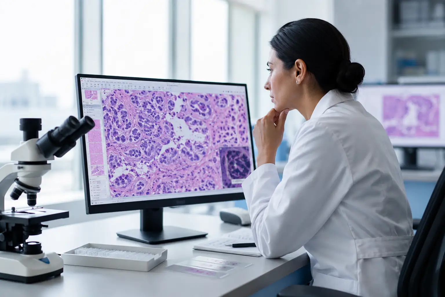
In many cases, a biopsy and pathology assessment are needed to establish a diagnosis. Imaging and tissue analysis answer different questions and are often used together. [2]
Why Specialist Interpretation Still Matters
Better images do not explain themselves. Radiologists assess the pattern of findings, compare earlier scans where available, and relate what they see to the clinical question. That interpretation gives the images their practical value.
When findings are complex or uncertain, an independent radiology second opinion may help clarify the report or confirm its interpretation. It does not guarantee a different diagnosis and should support discussion with the clinician responsible for ongoing care.
Hospitual offers a route to specialist review of existing imaging and reports. Patients can explore available specialists and select a doctor for their case. See how Hospitual works for the process.
Frequently Asked Questions
Which scan is best for detecting cancer?
There is no single best scan for every cancer. The choice depends on the suspected condition, the part of the body and the question the clinician needs to answer. Different scans may be used together.
Can a scan confirm cancer without a biopsy?
Sometimes imaging provides highly characteristic findings, but many diagnoses still require a biopsy. The treating team decides whether tissue confirmation or further tests are needed.
Can AI replace a radiologist?
AI can assist with defined tasks, but its output needs appropriate clinical oversight. A radiologist remains responsible for interpreting the examination within the wider clinical picture.
Key Takeaways
- X-rays, ultrasound, CT, MRI and PET provide different kinds of information.
- No scan can answer every clinical question or rule out every cancer.
- AI offers support for specific tasks, with validation and oversight.
- Specialist interpretation connects imaging findings to patient care.
References
- Sharma B, Martin A, Stanway S, Johnston SRD, Constantinidou A. Imaging in oncology – over a century of advances. Nature Reviews Clinical Oncology. 2012;9:728–737. doi:10.1038/nrclinonc.2012.195.
- National Cancer Institute. Tests and Procedures Used to Diagnose Cancer.
- National Cancer Institute. Computed Tomography (CT) Scans and Cancer.
- RadiologyInfo.org (ACR and RSNA). PET/CT – Positron Emission Tomography/Computed Tomography.
- National Cancer Institute. Can Artificial Intelligence Help See Cancer in New Ways? 2022.
Educational information only. This article does not provide an individual diagnosis or treatment recommendation. Discuss imaging results and next steps with your treating clinician.